Cornea Diseases
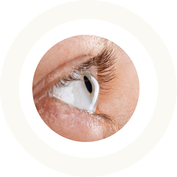
What is corneal disease?
The cornea is a transparent structure at the front of the eye, essential for clear vision. Corneal diseases can affect its different layers—epithelium, stroma, and endothelium—leading to reduced visual quality, discomfort, or severe complications.
Timely and specialized care is critical to preserving eye health and improving patients’ quality of life.
When to seek consultation:
symptoms of corneal diseases
Recognizing the symptoms of corneal diseases is essential to seek prompt specialist care.
If these symptoms occur, an ophthalmological consultation can enable early diagnosis and appropriate treatment.
Book an appointment today to discuss your options with one of our specialists.
Key symptoms include but are not limited to the following:
- Blurred or distorted vision
- Increased sensitivity to light (photophobia)
- Redness and irritation of the eye
- Persistent foreign body sensation or discomfort
- Frequent changes in eyeglasses or contact lens prescriptions

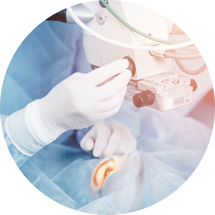
Types of corneal diseases and their treatments
Keratoconus
Keratoconus is a progressive eye condition in which the cornea, the transparent front part of the eye, thins and gradually bulges outward into a cone-like shape. This irregular shape leads to distorted vision, increased sensitivity to light, and frequent prescription changes for glasses or contact lenses.
This relatively common condition often begins in adolescence or early adulthood, with symptoms worsening over time. While the exact cause is unknown, genetic and environmental factors may play a role. Left untreated, keratoconus can cause significant vision loss, underscoring the importance of early diagnosis and management. Today’s treatments can halt the progression, which is why early diagnosis is key in maintaining good vision.
We have developed an app to help follow this disease, and have the most up-to-date technology to follow your keratoconus. The Anterion, an OCT based imaging device using the most modern algorithms, detects advancement in your condition. Along with the Anterion, our application also helps graphs and recognizes any changes, and reminds you of appointments as needed.
Treatment options for keratoconus
Several treatment options are available for keratoconus, depending on the stage of the disease and the severity of corneal changes. Each option is tailored to the patient’s specific needs, the degree of disease progression, and the overall condition of the cornea.
Glasses and contact lenses
In early stages, glasses or specially designed contact lenses can correct vision problems. Soft lenses may suffice initially, but rigid gas-permeable (RGP), hybrid lenses or scleral lenses may be needed as the condition progresses.
Corneal cross-linking (CXL)
This innovative procedure halts keratoconus progression by strengthening the corneal tissue. Riboflavin (vitamin B2) drops are applied to the eye and activated with ultraviolet (UV) light, creating new collagen cross-links to stabilize the cornea and prevent further thinning.
Laser treatments can be applied to improve vision at the time of this treatment in a private centre. Ask your physician if this is of interest to you.
Corneal transplantation
In advanced cases of keratoconus, when other treatments are no longer effective, a corneal transplant may be necessary. Different types of transplants are available depending on the severity of the disease:
Penetrating keratoplasty (PKP)
This technique involves replacing the entire central cornea with donor tissue. It is often used in severe cases where the cornea is extremely thin or scarred. While effective, this procedure requires several months of recovery and carries a small risk of graft rejection.
If this is the type of transplant that you have, ask about our app which can help follow cell counts in your graft as well as thickness, both parameters that help ensure a healthy transplant. Specular microscopy, a machine that counts endothelial cells, is available in our clinic. These measurements are part of our standard follow up for every corneal transplant.
Deep anterior lamellar keratoplasty (DALK)
This type of transplant replaces only the front layers of the cornea (mostly stoma), preserving the patient’s healthy endothelium. This reduces the risk of rejection and allows for faster visual recovery, although the procedure is technically more complex. Both Dr. Talajic and Dr. Choremis have access to a femtolaser, assisting in this procedure at Maisonneuve-Rosemont Hospital.
Dr. Choremis performed the first femtosecond laser-assisted corneal transplant in Quebec in March 2025 at Maisonneuve Rosemont Hospital’s Angus surgical centre. This pioneering technology will be accessible to both herself and Dr. Talajic moving forward, enhancing their capability to deliver cutting-edge treatments. At Haute Vision, our approach to managing keratoconus is both holistic and comprehensive. We tailor each treatment to the unique presentation of the disease in each patient, ensuring personalized and effective care. This adaptability and commitment to individualized treatment underscore our dedication to advancing keratoconus therapy.
Corneal allogenic intrastromal ring segments (CAIRS)
This innovative technique involves inserting donor ring segments into the cornea to reshape and stabilize its structure.
CAIRS offers an alternative to full corneal transplantation for certain patients with keratoconus, particularly in the early to moderate stages. This minimally invasive procedure helps to slow the progression of the disease and improve vision by regularizing the corneal shape.
In 2023, Dr. Julia Talajic was the first in Quebec to perform CAIRS, a novel treatment for keratoconus, with Dr. Johanna Choremis following soon after. Together, they established Haute Vision as a premier centre for this avant-garde therapy. Their holistic approach to keratoconus includes offering excimer-laser assisted PRK with Crosslinking, phakic IOL (EVO) implantation, CAIRS, or DALK/PK for keratoconus, depending on patient preferences and disease severity.
This procedure requires the use of a femtosecond laser, which is available to our patients at Maisonneuve-Rosemont Hospital, a renowned leader in eye care and advanced technology.
Endothelial diseases
Endothelial diseases primarily affect the innermost layer of the cornea, the endothelium, which regulates fluid balance and maintains corneal transparency. When endothelial cells are damaged or malfunction, the cornea can swell and become cloudy, resulting in blurred vision, glare, and discomfort.
The most common forms of endothelial diseases include Fuchs’ Endothelial Corneal Dystrophy (FECD) and Pseudophakic Bullous Keratopathy (PBK).
Fuchs’ endothelial corneal dystrophy (FECD)
Fuchs’ dystrophy is a progressive, hereditary condition where endothelial cells gradually deteriorate, causing guttata to appear in the deepest layers of the cornea, with eventual corneal swelling (edema). Over time, this causes glare and clouded vision. Eventually, swelling can lead to clouded vision, corneal scarring, and painful blisters (bullae) on the corneal surface. This can cause significant visual impairment.
Fuchs’ dystrophy typically becomes noticeable later in life, often after the age of 50, and is more common in women.
Ask your doctor about our app which can help follow Fuchs disease. We can incorporate the V Fuchs questionnaire, pachymetry, as well as specular microscopy counts, all parameters to help follow disease progression.
Pseudophakic bullous keratopathy (PBK)
PBK occurs when the corneal endothelium is damaged after intraocular surgery, such as cataract surgery, glaucoma surgery, or retinal surgery. This condition can cause painful swelling and blistering of the cornea. PBK is one of the leading causes requiring corneal transplantation in patients who have undergone cataract surgery, particularly in complex cases or those with pre-existing corneal conditions.
Treatment options for endothelial diseases
The most effective treatment for advanced endothelial diseases is corneal transplantation. Several types of transplants can be considered, depending on the extent of the endothelial damage and the overall health of the patient’s eye.
Corneal transplant rejection
Any patient that has a corneal transplant should be aware of signs and symptoms of a rejection. Any reduced vision, light sensitivity, redness or change in your eye can be a rejection after corneal transplantation. Seek medical attention immediately to be seen. Early treatment of corneal transplant rejection can reverse most rejections.
Our app is a valuable tool for any transplant involving endothelial replacement. It tracks cell counts in your graft and monitors its thickness—two critical parameters for maintaining a healthy transplant. Our clinic is equipped with specular microscopy, a specialized device for counting endothelial cells. These measurements are an integral part of our standard follow-up care for every corneal transplant.
Descemet Membrane Endothelial Keratoplasty (DMEK):
This revolutionary technique replaces only the damaged endothelial layer with a thin donor graft, preserving the rest of the cornea. DMEK provides numerous advantages, including exceptional success rates, faster recovery, fewer complications, and more precise visual outcomes. For advanced cases of Fuchs’ dystrophy, Descemet Membrane Endothelial Keratoplasty (DMEK) remains the gold standard, offering superior visual outcomes and the lowest risk of rejection.
Dr. Julia Talajic was the first ophthalmologist to perform DMEK in Quebec nearly a decade ago, setting a new standard in corneal surgery. Dr. Johanna Choremis followed, performing the first DMEK procedures at the Jewish General Hospital. Haute Vision is a leading centre for this innovative treatment.
Descemet membrane automated endothelial keratoplasty (DSAEK)
DSAEK is another partial thickness endothelial transplant that may be preferred in certain situations. For example, after glaucoma surgery, or following complex cataract surgery.
Penetrating keratoplasty (PKP):
In cases where more extensive damage to the cornea is present, PKP, or a full-thickness corneal transplant, may be required. PKP replaces the entire central cornea but involves a longer recovery period and a slightly higher risk of rejection.
Basement membrane dystrophy
Also known as Epithelial Basement Membrane Dystrophy (EBMD) or Map-Dot-Fingerprint Dystrophy, this condition affects the cornea’s outermost layer (epithelium), causing abnormal growth patterns. Symptoms may include recurrent corneal erosions, blurred vision, and ocular discomfort.
Treatment options for basement membrane dystrophy
Treatments include lubricating eye drops, ointments, and, in advanced cases, keratectomy, whether manual or with the aid of an excimer laser (Phototherapeutic Keratectomy—PTK). This procedure smooths the corneal surface to reduce symptoms.
Salzmann’s nodular degeneration
Salzmann’s nodular degeneration is a corneal condition characterized by bluish-white nodules forming on the corneal surface. These nodules can cause blurred vision and irritation. The condition is often associated with chronic surface inflammation, such as dry eye syndrome or blepharitis.
Treatment options for Salzmann’s nodular degeneration
Topical anti-inflammatory medications can alleviate symptoms, while surgical removal of the nodules may be required in advanced cases to restore corneal clarity.
Pterygia
A pterygium is a benign growth of fleshy tissue that begins on the sclera (white part of the eye) and can extend onto the cornea, potentially affecting vision. This condition is more common in individuals exposed to high levels of UV light, dust, or wind. It is sometimes referred to as “Surfer’s eye.” Common symptoms include redness, irritation, and a sensation of a foreign body in the eye. If it impinges far into the cornea, it can create distortion in the vision, usually in the form of astigmatism.
Treatment options for pterygia
Small pterygia can be managed with lubricating eye drops to reduce discomfort. However, larger pterygia that impair vision, induce astigmatism, or cause significant discomfort may require surgical removal. During surgical removal, conjunctival autografts are often performed. This means a small piece of healthy conjunctiva (clear membrane over the white of the eye) is taken to cover the defect created by the surgery. When this technique is not used, recurrence rates can be as high as 50%. Our team systematically performs these grafts during pterygium excision using physiologic glue (+/- sutures) to reduce inflammation, reduce discomfort and minimize recurrences.
TOP CHOICE FOR SPECIALIZED EYE CARE
Why choose Haute Vision
for corneal diseases treatment?
Expertise in corneal surgery
Our surgeons are recognized leaders in the field of corneal diseases. Their expertise spans a wide range of treatments, from innovative medical options to complex surgical procedures.
State-of-the-art technology
We use advanced diagnostic tools and cutting-edge surgical techniques, such as DMEK and CAIRS, to ensure precise and effective care tailored to the specific needs of each patient.
Personalized treatment plans
Every treatment plan is customized based on the patient’s condition and expectations. Whether addressing keratoconus, Fuchs’ dystrophy, or another corneal disease, we provide solutions designed to optimize visual outcomes and enhance quality of life.
Comprehensive follow-up care
Our approach extends beyond surgical procedures. We offer thorough follow-up care to monitor the disease progression and corneal health while managing any potential complications to ensure long-lasting results. Ask about our app to help follow many of these corneal diseases.